After skin wrinkles and sagging skin, the next most common skin concerns I see in the office are darkened (hyperpigmented) skin patches and growths, and also vascular skin blemishes (i.e. telangiectasias and varicosities). Let’s look closer at how to treat these.
Hyperpigmentation
The sun’s ultraviolet radiation triggers cells in the basal skin layer to produce melanin. These cells are called melanocytes, and the skin pigment they produce is generally considered your body’s natural protection against sun-induced damage.
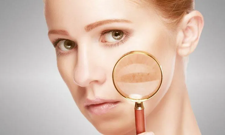
Hyperpigmented spots and growths come in varying sizes and configurations: Lentigines, solar lentigo, or “age spots” are flat dark skin patches Ephelides, or “freckles” are small dark spots Melasma, chloasma, or “mask of pregnancy” are large symmetrical face patches Café au lait spots are isolated flat larger dark spots Actinic keratoses, or solar keratoses, or “senile keratoses” are raised thick, scaly, or crusty benign skin lesions with varying pigments: dark, tan, pink, red, or lightened.
The simplest approach to lightening your skin spots is by using a topical cream containing hydroquinone 2-4% or kojic acid. This is best if you have one or just a few small brown patches. My local compounding pharmacy has “Dr. Cutler’s skin-lightening cream,” which I have personally used and achieved an excellent result in just one month of nightly application.
It contains the following ingredients: tretinoin (e.g. Retin-A) 0.025%, mometasone furoate 0.1% (a mid-potency steroid), and hydroquinone 4%. Any compounding pharmacy can do the same for you, with a prescription from your doctor. There are other topical creams that can lighten skin, but much more slowly.
For example, niacinamide (vitamin B3) 5% topical was shown to reduce hyperpigmentation (age spots and melasma), red blotchiness, and wrinkles in a double-blind placebo- controlled split face study of women ages 40 to 60 years old followed over twelve weeks.
1. Other studies repeatedly prove it has anti-itch, antimicrobial, photo-protective, anti-oil and color-lightening effects depending on its concentration.
2 You can achieve faster results for larger areas of skin and skin growths that need lightening or destruction by using intense pulsed light (IPL) therapy. Let me tell you what I’ve learned about this.
Intense Pulsed Light (IPL) is much like LASER light therapy, but more comfortable. IPL treats uneven skin tones, blotchy appearances, dark-colored patches, and much more.
It is fascinating how it works so safely and effectively. Let me explain it. Intense Pulsed Light (IPL) is a broadband of multiple light wavelengths (versus LASER in which light waves are synchronized). IPL technology and LASER are based on the principle of selective photothermolysis: photo (light)—thermo (heat)—lysis (destruction).
The intense pulse of light is sent through a filter that is designed for specific light- sensitive molecules of the skin, called “chromophores.” The filter is needed to convert the light to a desired wavelength that can heat up and destroy only the “target” chromophore, and not the surrounding tissue.
In this case, the chromophore is melanin (the molecule of skin pigment). The light therefore, causes a photochemical heat reaction only at the melanin (pigment) molecules. It’s quite ingenious if you ask me. For example, one of the newest and most effective treatments on the market is the Viora V-20 (www.vioramed.com) which is exclusively designed to safely, effectively and rapidly reduce melanin in the skin surface layers for visibly lighter and more even skin tones.
This is what I use in my office. The Viora has different filters needed for the specific target tissue type (e.g. hair pigment, skin pigment, hemoglobin molecule of blood vessels, etc.) and also for the specific skin tone type (including all Fitzpatrick skin types 1 through 6). Treatments are further fine-tuned by a range of the following parameters: energy intensity (Joules) of the light flash light pulse configuration (single, multiple, or rapid) light pulse duration relaxation time between pulses Safety and comfort are enhanced by Viora’s built-in thermal electric cooling system.
If needed, topical numbing cream can be applied for comfort. Vascular lesions Tiny blood vessels in skin create the appearance of the varying vascular lesion types: Hemangiomas: excess dense blood vessels appear rubbery or like a bright red nodule Telangiectasias: tiny superficial red, blue or purple arterioles or venules are most often seen on the cheeks, nose, chin, neck, chest or spider veins on legs Rosacea and couperose: confluent redness of the skin due to small dilated blood vessels.
Rosacea is an acne-like inflammatory condition affecting more than 14 million people Hematoma (bruising) usually from trauma or disease Poikiloderma: skin with a mixed appearance of hyperpigmentation, hypopigmentation, telangiectasias and atrophy usually on the chest or the neck, due to sun damage Port wine stains: seen in 3 out of every 1,000 children born, this is a large flat red pigmented skin area appearing as a skin “stain” under the skin. I don’t know of any topical creams that can effectively destroy vascular lesions. Therefore, IPL is my preferred method. With vascular lesions, the target chromophore is hemoglobin (the pigment molecule in red blood cells that carries oxygen). The filter for the IPL therefore causes heat energy to directly coagulate and destroy only the blood vessels without damaging the surrounding tissue. The damaged tiny blood vessels are then broken down by your natural enzymes and tissue healing mechanisms, and gradually disappear over many days to weeks.
To long-term health and feeling good,
Michael Cutler, M.D.


